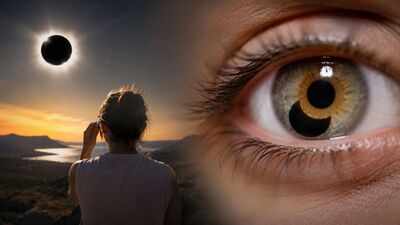
On the afternoon of 21 August 2017, millions of people across the United States looked towards the Sun as the Moon moved across it. For most viewers, the eclipse became a short-lived memory once the sky returned to normal. For Nia Payne, a 26-year-old woman in Staten Island, the event left something behind. Within days, she noticed a small disturbance in the centre of her vision. It was not simply a blur or a temporary after-image. The shape was recognisable. It looked like a crescent, almost as if part of the eclipse had remained in her sight after she had stopped watching it. Doctors would later discover that the unusual shape corresponded to actual damage inside her retina.
2017 solar eclipse left a woman with crescent-shaped blind spot
Payne was watching a partial eclipse from New York, where around 70%of the Sun was covered by the Moon. Unlike observers inside the narrow path of totality, she never saw the Sun completely disappear. She did not have suitable eclipse glasses when she first looked up. According to the case later published by ophthalmologists at Mount Sinai, she stared at the Sun for approximately six seconds before looking away.She then borrowed a pair of glasses from someone nearby and looked again. The glasses were later found not to meet the ISO 12312-2 standard for safe solar viewing. This second period of viewing lasted somewhere between 15 and 20 seconds.By the time she went indoors, Payne had begun to notice a blurred patch in the centre of her left eye. It did not disappear over the next day or two. Instead, the disturbance became more clearly defined. The shape occupied the central part of her vision, which meant that it appeared over whatever she was trying to look at. Reading, focusing on objects and recognising details could all become difficult when they fell within that area.
The problem was deeper than ordinary blurred vision
According to The Washington Post, several days after the eclipse, Payne was examined at the New York Eye and Ear Infirmary of Mount Sinai. Her standard visual acuity measurements did not initially suggest a devastating injury. Her right eye tested at 20/20, while her left eye measured 20/25. Those figures were relatively close to normal, but they did not capture the problem she was experiencing.The difficulty was not that she had suddenly lost all of her sight. Instead, a small but important part of her central visual field had been damaged. Doctors examining her retina found evidence consistent with solar retinopathy. The condition occurs when direct sunlight causes injury to the light-sensitive tissue at the back of the eye.The retina is a thin layer of specialised nerve tissue lining the inside of the eye. It contains photoreceptors that respond to light and help convert what the eye sees into signals that can be processed by the brain. Among these cells are rods and cones. Rods are particularly important for vision in low light, while cones provide much of the detailed and colour vision used during daylight. The highest concentration of cones is found around the fovea, a tiny central region responsible for sharp vision. That was significant in Payne’s case because the damage had occurred in an area involved in central vision.
What happens to the retina during solar retinopathy
The eye is remarkably effective at gathering light. Its lens focuses incoming rays onto the retina, allowing a person to see fine details with considerable precision.That same optical system becomes dangerous when the source is the Sun. Direct sunlight can be concentrated onto a very small area of retinal tissue. The resulting injury can involve both heat-related effects and photochemical damage. High-energy light can trigger chemical reactions within and around photoreceptor cells, while intense illumination can also contribute to thermal injury.The retina is particularly vulnerable because it is not designed to withstand prolonged exposure to concentrated sunlight. A person may not feel an immediate, severe warning from the retina itself. The absence of obvious pain does not mean that the tissue is unharmed.Once photoreceptor cells have been destroyed, the body cannot simply replace them with new cells. This is one reason solar retinopathy can leave a lasting blind spot.
Doctors used retinal imaging to trace the crescent-shaped damage
One of the most striking details of Payne’s examination came from something very simple. The doctors asked her to draw the area she could no longer see properly.Rather than trying to describe the disturbance with medical terminology, she put the shape on paper. The result was a crescent corresponding to the area missing from her central vision.The drawing was important because the shape was not random. It resembled the portion of the Sun that had remained visible during the partial eclipse. They used adaptive optics scanning light ophthalmoscopy, an advanced imaging technique capable of producing extremely detailed views of retinal structures. Adaptive optics was originally developed for astronomy, where it is used to compensate for distortions caused by Earth’s atmosphere.In ophthalmology, the same principle can be applied to correct for small imperfections in the eye’s own optical system. This allows clinicians to examine individual photoreceptors and other microscopic structures in a living eye. The images taken from Payne’s eye showed areas of photoreceptor damage.
The image inside her eye matched the eclipse
According to the study published in JAMA Ophthalmology, titled ‘Acute Solar Retinopathy Imaged With Adaptive Optics, Optical Coherence Tomography Angiography, and En Face Optical Coherence Tomography’ reveals the scans showed a region of photoreceptor disruption in Payne’s left eye and a less pronounced pattern in her right eye. The orientation and shape of the damage corresponded to the crescent she had reported seeing.When Payne looked towards the partially eclipsed Sun, the eye’s lens focused the incoming light onto the retina. The shape of the light reaching the retina reflected the shape of the visible portion of the Sun.The exposed photoreceptors were therefore damaged in a pattern that broadly followed that shape. The result was an unusual chain of events in which the object she had looked at became, in effect, a map of the injury it caused.Her drawing represented what she could no longer see clearly. The retinal scan showed where the cells had been damaged. Both followed the geometry of the eclipse.
Why a partial solar eclipse can still damage your eyes
The case also illustrates why an eclipse does not make direct sunlight safe to look at. During totality, when the Moon completely covers the bright surface of the Sun, there is a brief period when direct viewing can be safe under specific circumstances. That period ends as soon as the bright solar surface begins to reappear.A partial eclipse is different. The Sun remains visible throughout the event, even though part of its disc is hidden.In New York, Payne was watching a partial eclipse rather than totality. Most of the Sun was covered, but a substantial portion remained exposed. Ordinary sunglasses do not provide the protection required for direct solar viewing. Properly manufactured eclipse viewers and solar filters are designed to reduce the Sun’s radiation to safe levels and should meet the relevant safety standard.The distinction matters because the eye’s normal response to bright light is not a reliable protection against retinal damage.
The damage remained after the eclipse had passed
Payne’s case was followed because solar retinopathy does not have a simple treatment that can restore destroyed photoreceptors.Some people with solar retinopathy experience partial improvement in their vision after the initial injury. In other cases, the central disturbance can remain. For Payne, the crescent-shaped area continued to interfere with central vision. She adapted by relying more heavily on her right eye, but reading and looking at screens could still be difficult.The problem was small in physical size but significant in everyday use because it occupied the centre of her field of view. A person can compensate for many forms of peripheral visual loss without constantly noticing them. A defect directly in the area used to read words or examine a face is much harder to ignore.
How the eclipse left its shape on her vision
The Mount Sinai case became unusual not simply because Payne developed solar retinopathy, but because doctors were able to see the injury at such fine resolution and compare it with what she reported seeing. Her drawing provided a visual record of the blind spot. The adaptive optics images provided a record of the damaged photoreceptors.What happened was not that an actual picture of the eclipse was literally stored inside the eye. The effect was more precise and more biological than that. The eye’s optical system projected the partially obscured Sun onto the retina, and the intense light damaged the photoreceptors receiving that image.The surviving cells around the damaged area continued to function, while the injured region produced a corresponding disturbance in her visual field.